Research-Dedicated Human Imaging Suites
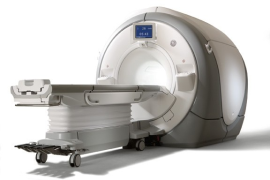
The Brain Imaging and Analysis Center (BIAC) at Duke University houses two GE UltraHigh Performance (UHP) 3.0 Tesla (T) scanners, offering a unified platform and enabling parallel access for research-dedicated human imaging. Through a productive collaboration with the GE Healthcare, both scanners are running on the latest GE UHP platform based on the GE Premier system architecture. Compared to the standard Premier products, our two systems have upgraded 60-cm high-power torque-balanced gradient coils that have gradient strengths at 100 mT/m (constant mode) and 113 mT/m (burst mode) with a slew rate of 250 T/m/s. On the RF side, our systems are equipped with 128ch receiver chain and the integrated local RF/Bo shimming capabilities complimenting the onboard whole-body high-order shimming coils. In addition to this advanced hardware, our two 3T scanners also have the latest pulse sequences, including multi-band, compressed sensing, and ultrahigh-resolution imaging as standard features. Finally, both 3T scanners are connected to our supercomputer and solid-state storage server via a high-speed network interface.
Each human scanner suite is equipped with Resonance Technology 3D TV and/or projectors for visual stimulation. Both scanners have integrated eye tracking capability and MR-compatible auditory systems. Additional sensory stimulation equipment includes a Mindware Technologies olfactometer, a Medoc thermode system, eye-tracking software, BIO electrodes for galvanic skin response recording, and a Grass Instrument’s constant current nerve stimulator. Subject responses are obtained from 4-button and 8-button boxes and joysticks. Each suite is equipped with a pair of high-performance PCs equipped with multi-function boards for timing, digital I/O, and A/D conversion that maintains experimental control in each scanner suite. A variety of software packages are available for experimental control, including E-Prime, Presentation, and MATLAB PsychToolbox.
An MRI-compatible physiological monitoring system (In-Vivo Research) provides continuous measurement of ECG, end-tidal CO2, respiration, and non-invasive blood pressure. This system is used during clinical studies involving sedation, or studies involving at-risk individuals.
MRI Simulation Facilities

To acclimate patients to the MRI scanner experience, BIAC has recently purchased a new Vera MRI Simulator from Psychology Software Tools to replace the old mock scanner used in previous years. This new simulator includes an automated table, lights and fan, and provides an improved platform for potential subjects to experience what it will be like in the actual MRI machine. While the participant is in the mock scanner, you can display tasks to the subject, as well as play MRI sounds for various sequences.
This system is a great resource to introduce participants to the identical experimental procedures that they will experience in the actual scanner, and to train children to participate in functional neuroimaging studies. This is done to ensure participant comfort and data quality.
For additional operating tips and procedures, please visit https://wiki.biac.duke.edu/biac:scanners