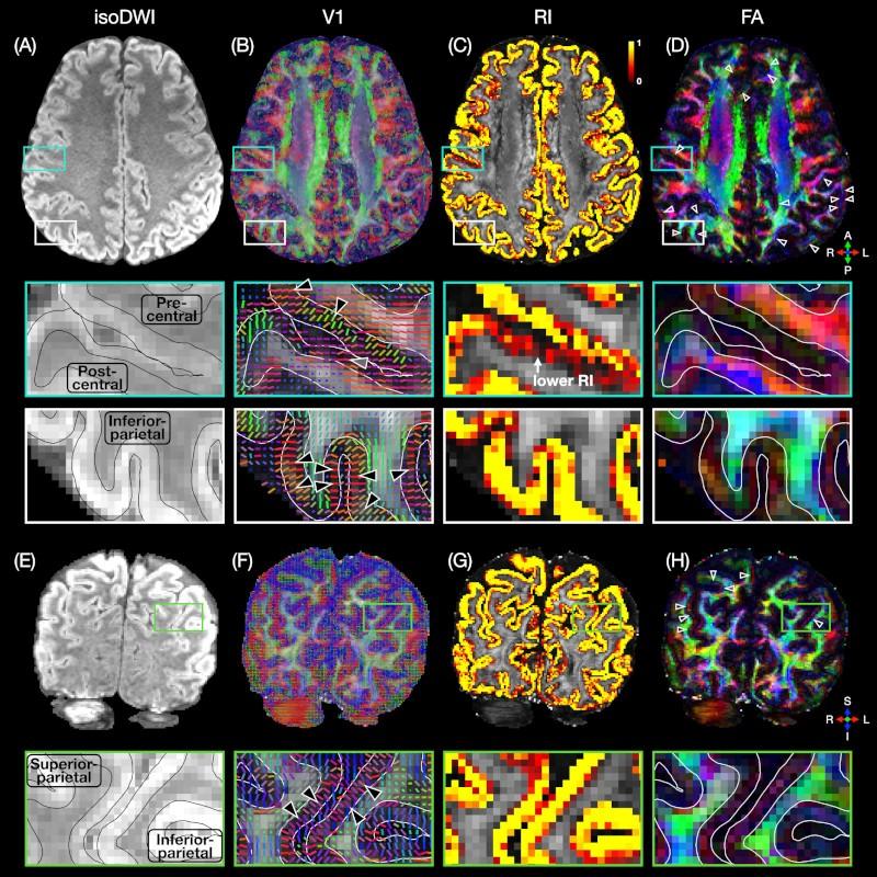
High-resolution diffusion tensor imaging (DTI) can noninvasively probe the microstructure of cortical gray matter in vivo. In this study, 0.9-mm isotropic whole-brain DTI data were acquired in healthy subjects with an efficient multi-band multi-shot echo-planar imaging sequence. A column-based analysis that samples the fractional anisotropy (FA) and radiality index (RI) along radially oriented cortical columns was then performed to quantitatively analyze the FA and RI dependence on the cortical depth, cortical region, cortical curvature, and cortical thickness across the whole brain, which has not been simultaneously and systematically investigated in previous studies. The results showed characteristic FA and RI vs. cortical depth profiles, with an FA local maximum and minimum (or two inflection points) and a single RI maximum at intermediate cortical depths in most cortical regions, except for the postcentral gyrus where no FA peaks and a lower RI were observed. These results were consistent between repeated scans from the same subjects and across different subjects. They were also dependent on the cortical curvature and cortical thickness in that the characteristic FA and RI peaks were more pronounced i) at the banks than at the crown of gyri or at the fundus of sulci and ii) as the cortical thickness increases. This methodology can help characterize variations in microstructure along the cortical depth and across the whole brain in vivo, potentially providing quantitative biomarkers for neurological disorders.
PubMed link: https://pubmed.ncbi.nlm.nih.gov/36863550/
Citation: Ma Y, Bruce IP, Yeh CH, Petrella JR, Song AW, Truong TK. Column-based cortical depth analysis of the diffusion anisotropy and radiality in submillimeter whole-brain diffusion tensor imaging of the human cortical gray matter in vivo. NeuroImage. 2023;270:119993. doi: 10.1016/j.neuroimage.2023.119993