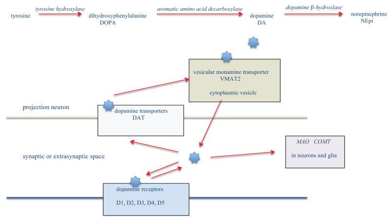
Advances in molecular and structural and functional neuroimaging are rapidly expanding the complexity of neurobiological understanding of Parkinson's disease (PD). This review article begins with an introduction to PD neurobiology as a foundation for interpreting neuroimaging findings that may further lead to more integrated and comprehensive understanding of PD. Diverse areas of PD neuroimaging are then reviewed and summarized, including positron emission tomography, single photon emission computed tomography, magnetic resonance spectroscopy and imaging, transcranial sonography, magnetoencephalography, and multimodal imaging, with focus on human studies published over the last five years. These included studies on differential diagnosis, co-morbidity, genetic and prodromal PD, and treatments from L-DOPA to brain stimulation approaches, transplantation and gene therapies. Overall, neuroimaging has shown that PD is a neurodegenerative disorder involving many neurotransmitters, brain regions, structural and functional connections, and neurocognitive systems. A broad neurobiological understanding of PD will be essential for translational efforts to develop better treatments and preventive strategies. Many questions remain and we conclude with some suggestions for future directions of neuroimaging of PD.
Citation: Weingarten, C. P., Sundman, M. H., Hickey, P., & Chen, N. K. (2015). Neuroimaging of Parkinson's disease: Expanding views. Neuroscience and biobehavioral reviews, 59, 16–52. https://doi.org/10.1016/j.neubiorev.2015.09.007